
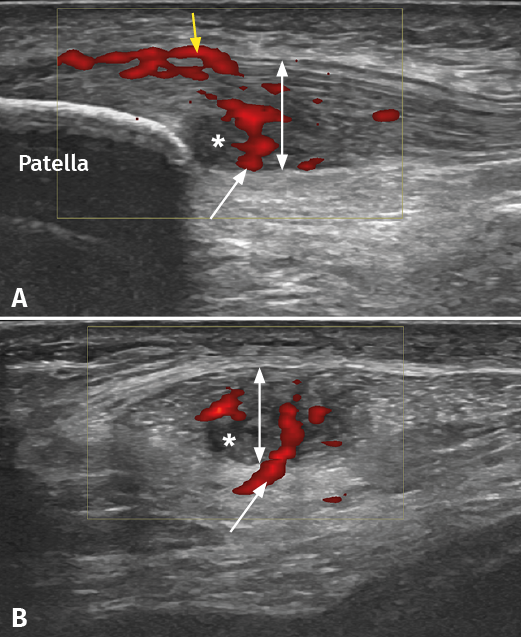
Figure 2. Ultrasound study of proximal patellar tendinopathy. A: longitudinal view of the patellar tendon; B: cross-sectional view of the same tendon. The white arrow shows hypervascularization from Hoffa's fat pad, while the yellow arrow indicates the increase in vascularization at surface (bursa) level. The double white arrow shows the increase in tendon thickness accompanied by hypoechogenic tissue degeneration zones (asterisk).