
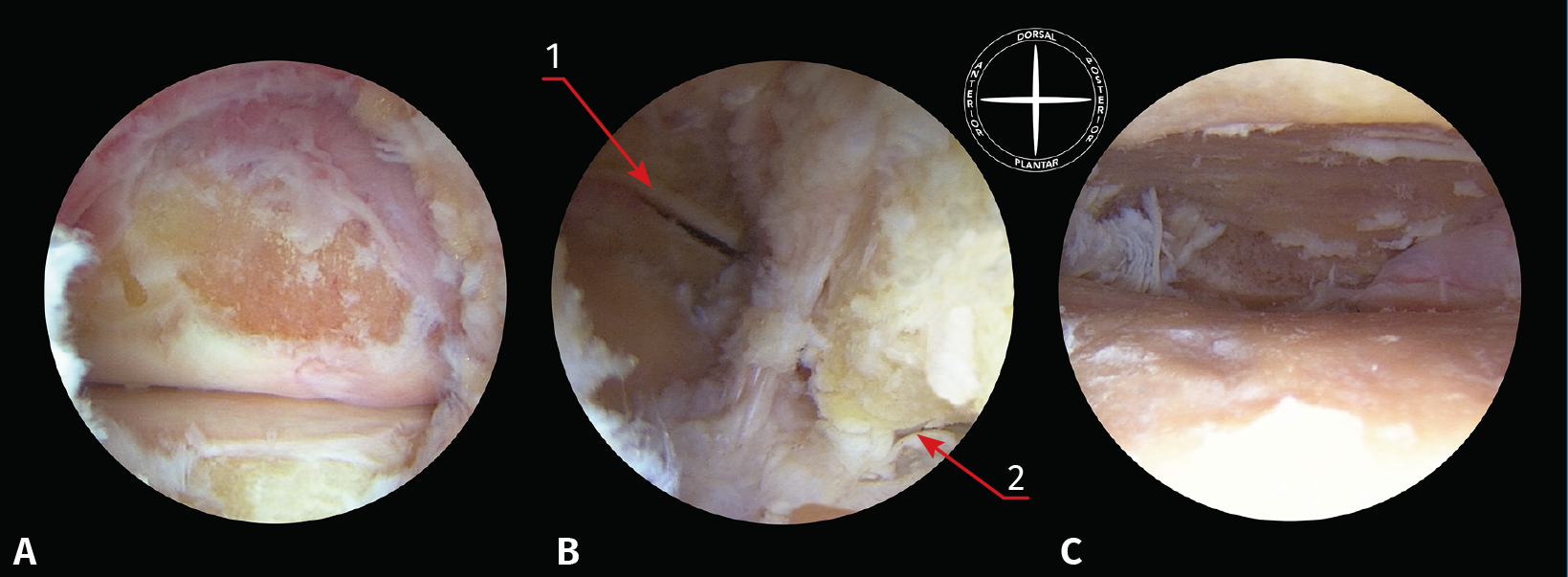
Figure 4. Different steps of the lateral portals or tarsal sinus arthrodesis technique (left ankle in supine decubitus with viewing through the anterolateral portal). A: visualization of the posterior subtalar joint after synovectomy; B: the same view after debridement of the capsule and fibrous tissue, exposing the cervical ligament of the tarsal sinus; C: the same view after completing joint cartilage denudation. 1: fibular malleolus; 2: fibular trochlea.