
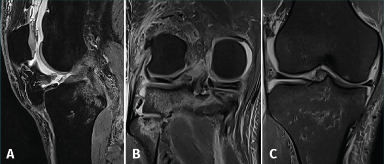
Figure 3. Magnetic resonance imaging study showing post-contusion edema of the tibia, with rupture of the anterior cruciate ligament. The posterior cruciate ligament appears correctly inserted in the sagittal view (A); the coronal view shows a radial lesion of the external meniscal root and bone avulsion with a bone fragment of the external root (B), causing extrusion of the internal meniscus (C).