
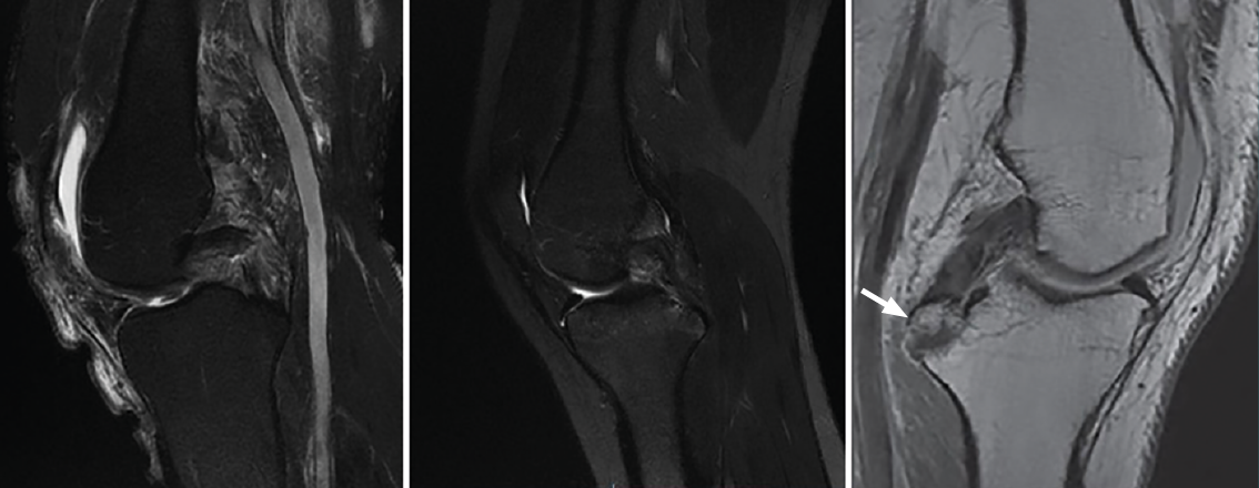
Figure 6. A: magnetic resonance imaging (MRI) sagittal view in T2-weighted sequencing, showing partial rupture; B: complete rupture of the posterior cruciate ligament (PCL); C: MRI sagittal view in T1-weighted sequencing of the left knee. The white arrow indicates avulsion of the PCL with a bone fragment in the tibial insertion.