
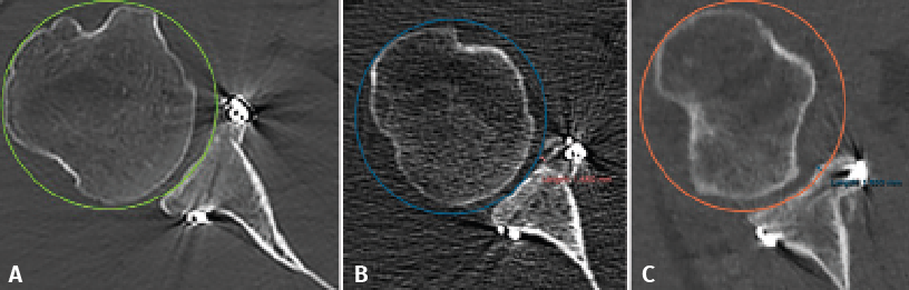
Figure 3. Evaluation of the graft position in the axial view, according to the description of Kany et al.(14). A circumference is traced over the glenoid border, and following the curvature of the latter, the amount of graft (in mm) extending beyond or failing to reach this line is measured. Image A shows the graft perfectly positioned, following the curvature of the glenoid concavity. Image B shows the graft medial with respect to the line. Image C shows the graft positioned too lateral.