
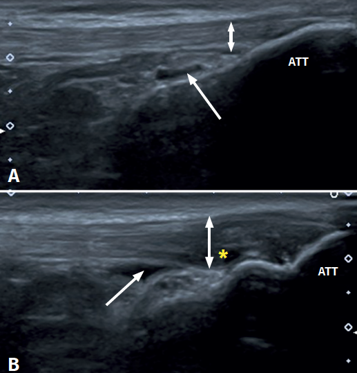
Figure 1. Ultrasound images (linear probe) of the patellar tendon, distal portion. Longitudinal view of the tendon fibres at insertion in the anterior tibial tuberosity (ATT). A: distal insertion of the normal patellar tendon. The arrow shows mild bursitis, and the double arrow indicates the normal size of the tendon; B: distal patellar tendinopathy showing the deep infrapatellar bursa (arrow) containing fluid (hypoechoic image), with increased tendon thickness (double arrow) and a fibrillar pattern showing decreased echogenicity (hypoechoic) secondary to disruption of the collagen bands (asterisk).