
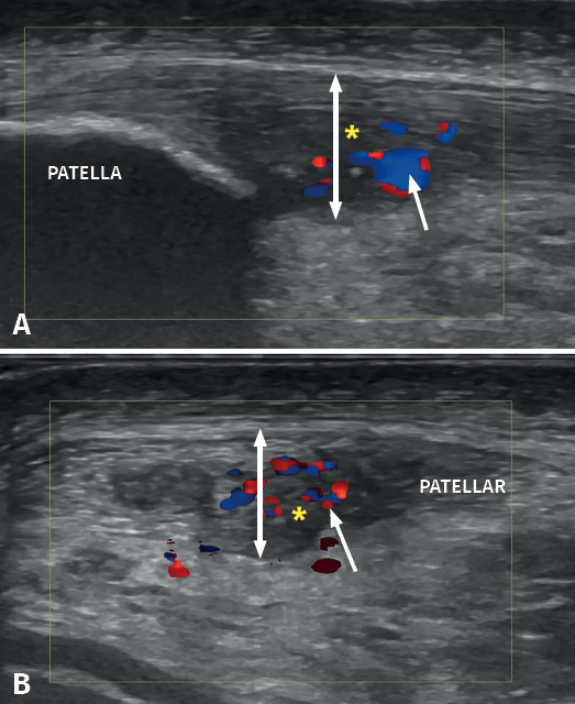
Figure 6. Doppler ultrasound in a patient with patellar tendinosis. The image A corresponds to a longitudinal view, while image B shows a cross-sectional view. The double arrow shows the increase in tendon thickness, while the asterisk indicates the hypoechogenic zones corresponding to the fibrillar lesion areas. The arrow shows hypovascularisation predominantly located in the deep and central zone.