
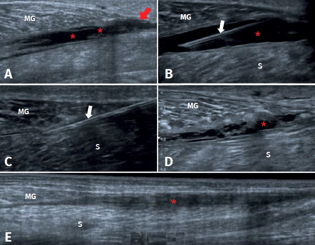
Figure 2. Evolution of a medial gastrocnemius fibrillar tear. A: ultrasound view of an acute tear of the distal myotendinous junction of the medial gastrocnemius (MG) (red arrow), associated to haematoma in the aponeurotic plane adjacent to the soleus (S) (asterisks); B: needle positioning before aspiration (white arrows) of the haematoma under ultrasound guidance; C: needle aspiration (white arrows) of the haematoma under ultrasound guidance, extraction of 70 cc; D: control after 10 days, haematoma relapse, of smaller size, with septa and signs of organization; E: control after 31 days, showing organization of the haematoma, evolving towards a fibrous scar cord, without signs of inflammation. Complete healing of the tear (red arrow).