
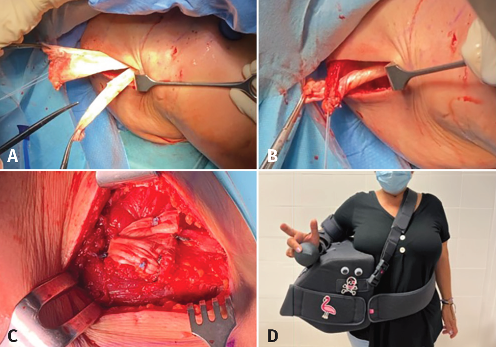
Figure 6. Right shoulder, patient placed in deck chair position, posterior view (A-C) and final view of the patient with postsurgical immobilization (D). A: a horizontal incision of the Achilles tendon graft is made at its free extremity; B-C: the free segments are interlinked one over the other to finally pass them through the loop of the lower trapezius, with fixation using non-resorbable sutures - thus connecting the Achilles tendon to the rest of the lower trapezius (LT) according to the Pulvertaft technique; D: final view of the patient with immobilization to maintain the arm in abduction and external rotation for 6 weeks.