
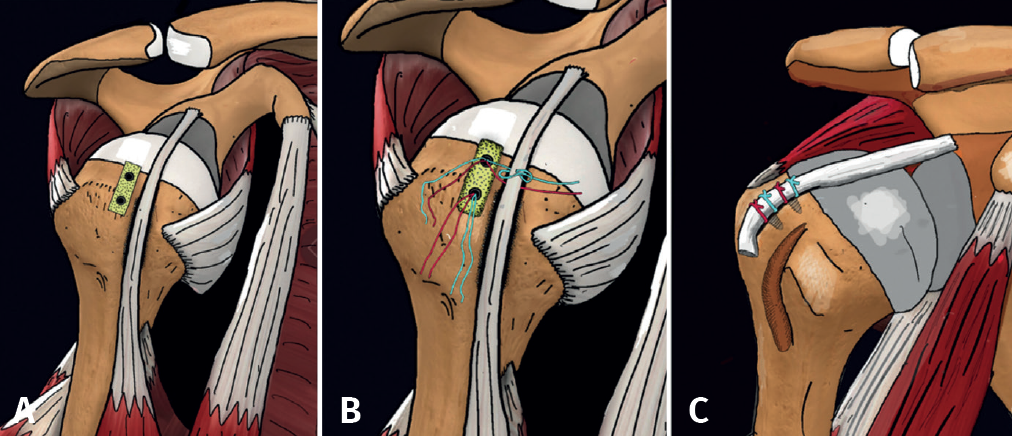
Figure 6. Right shoulder. A: preparation of the footprint parallel to the bicipital groove and decorticating part of the more lateral cartilage of the joint; B: appearance of the biceps after performing the two types of sutures with the threads of the more medial implant; C: final view of the technique after performing the biceps tenotomy. Reproduced from the original article by Llanos-Rodríguez et al.(56).