
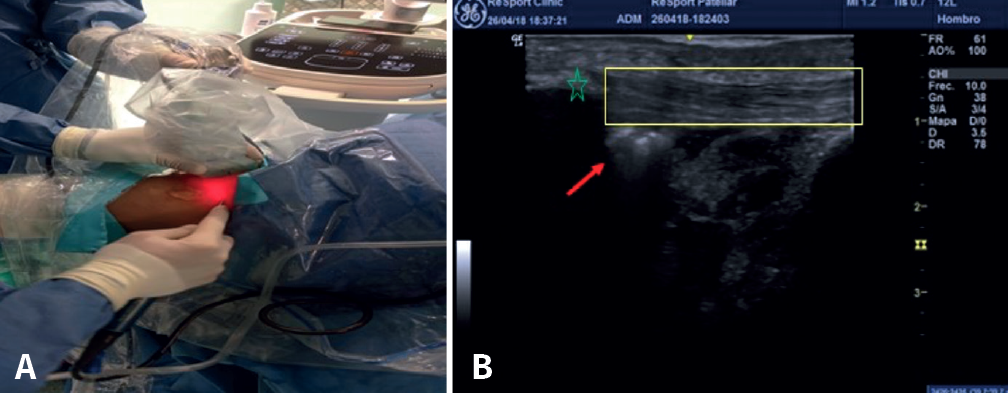
Figure 1. A: after creating the anterolateral and anteromedial knee portal 1 cm more lateral and more medial, respectively, resection is carried out of the neovessels and nerve tissue of the posterior aspect of the patellar tendon, as well as of the area of tendinosis and/or calcification in the posterior zone, until a tendon is obtained of the same thickness as the healthy contralateral tendon(74). The technique requires an assistant to hold the ultrasound device while the main surgeon controls the optics and the synoviotome. B: the red arrow indicates the location of the synoviotome over the deep zone of the patellar tendon (yellow inset). The asterisk shows the distal pole of the patella.