
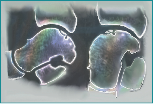
Figure 7. A preoperative CT scan performed with the ankle in forced plantarflexion can be a valuable tool in surgical planning. The sagittal image on the left reveals a posteromedially located osteochondral defect (OCD). In plantarflexion on the right, the same view illustrates the intraoperative position the surgeon will achieve. Two key anatomical changes occur: 1) A gap forms between the anterior distal tibia and the talus, improving access to the posterior joint space and the OCD; 2)The OCD shifts anteriorly. Using the anterior rim of the distal tibia as a reference easily identifiable intraoperatively.